GENERAL INFORMATION
Myxoma is a rare benign soft tissue tumor composed of fibroblasts embedded in abundant myxoid stroma. Myxomas have a gelatinous consistency, resembling the fetal umbilical cord. They may be derived from modified fibroblast cells. Sometimes, malignant tumors can undergo myxoid degeneration, making difficult to distinguish from myxomas on an MRI. In other words, malignant myxoid tumors can have a similar appearance as a myxoma on an MRI which is also similar to a ganglion cyst.
CLINICAL DATA
A benign soft tissue tumor
Usually solitary
Usually affects age 40 to 70 years
Slight female predilection
Multiple myxomas are rare
Multiples myxomas are associated with Carney complex:
Autosomal dominant disorder
Multiple cardiac and skin myxomas
Skin pigmentation
Endocrine involvement
Bone tumors
Mazabraud syndrome
Rare condition of multiple myxomas with polyostotic fibrous dysplasia
Multiple intramuscular myxomas localized in one side of the body
Polyostotic fibrous dysplasia appears before the myxomas
May be associated also with McCune-Albright syndrome
Three forms
Intramuscular myxoma (most common but still quite rarely seen)
Hypocellular
Sparse or inconspicuous vasculature
Bland spindle to stellate vasculature
Arises in skeletal muscle
Juxta-articular myxoma
Morphologically similar to intramuscular myxoma
Occurs immediately adjacent to a large joint
Commonly associated with cystic degeneration of adjacent articular cartilage
Thought to be related to a ganglion cyst
Digital myxoma
Digital lesion usually found in the finger
Composed of fibroblasts which elaborate a copious myxoid matrix
DIFFERENTIAL DIAGNOSIS
Low grade myxofibrosarcoma
Nerve sheath tumor
Angiomyxoma
Leiomyoma
Leiomyosarcoma
Ganglion Cyst
CLINICAL PRESENTATION
Signs/Symptoms
Intramuscular myxoma
Slowly growing
Painless intramuscular mass
Juxta-articular myxoma
Similar to intramuscular, near a big joint
Pain
Digital myxoma
Small
Painful
Prevalence
Digital and intramuscular myxoma more commonly affect females
Juxta-articular myxomas affect primarily males
Relatively uncommon
Affect primarily older adults (40-70 years of age)
Sites
Intramuscular myxoma
Thigh or gluteal region are most common sites
Also occur in the shoulder girdle and arm
Juxta-articular myxoma
Knee
Shoulder
Elbow
Hip
Digital myxomas are found on the finger
RADIOGRAPHIC PRESENTATION
Plain x-ray
Nonspecific soft tissue mass
Internal calcifications are rare
CT scan
Well-defined, homogenous soft tissue mass
Tissue attenuation, less than normal muscle
MRI
Well-defined mass
Homogenous signal
Intermediate signal intensity on T1W similar to muscle
High signal intensity on T2W indicative of fluid
Thin rim of higher signal around the mass on T1W
Post contrast heterogeneous enhancement on T1W
Enhance with contrast as opposed to a ganglion cyst
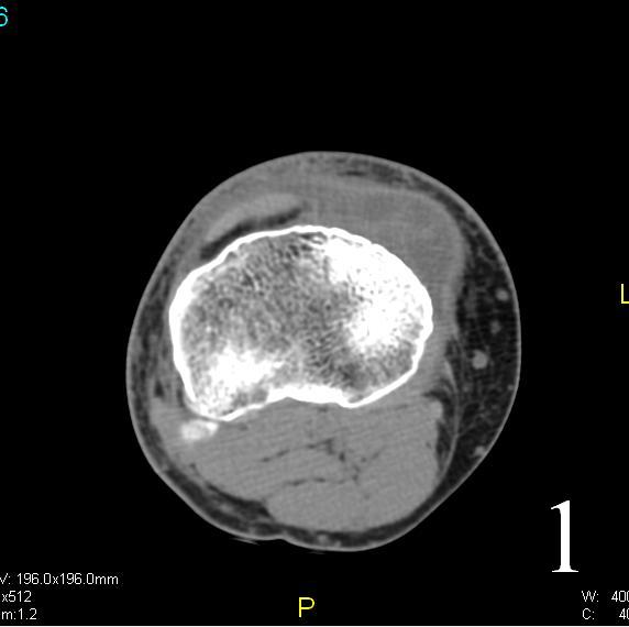 Fig. 1 Axial CT reconstruction of a myxoma of the knee, shows an anterior medial mass isointense with muscle.
%20of%20myxoma%20T1@0.jpg)
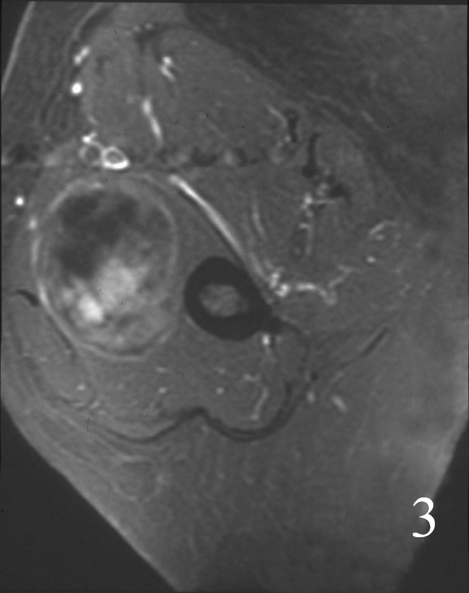
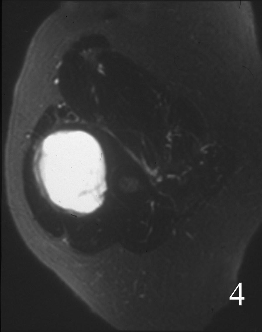 Fig. 2 Axial MRI of a myxoma of the upper extremity shows a homogeneous intermediate signal mass on T1W images (Fig. 2), heterogenous enhancement on post gadolinium (Fig. 3), and homogeneous high signal on T2W images (Fig. 4).
PATHOLOGY
Gross
Mucoid-gelatinous
Well circumscribed
Sometimes present with infiltrative borders
Rarely exceeds the 13 cm Ø
Microscopic
Intramuscular myxoma
Fairly well circumscribed but often infiltrates distally and proximally with what looks like a tail
Appears infiltrative
Merges with surrounding skeletal muscle and fascial tissue
Very hypocellular
Bland uniform hypocellular fibroblasts (spindle cells) in a mucinous bluish staining background
Juxta-articular myxoma
Morphologically identical to intramuscular myxoma
Digital myxoma
Similar feature to intramuscular
Hypercellulary is more common
Overlying epidermis may be ether hyperplastic or athrophic
Does not infiltrate into deeper tissues
Lesion merges imperceptibly with dermal connective tissue
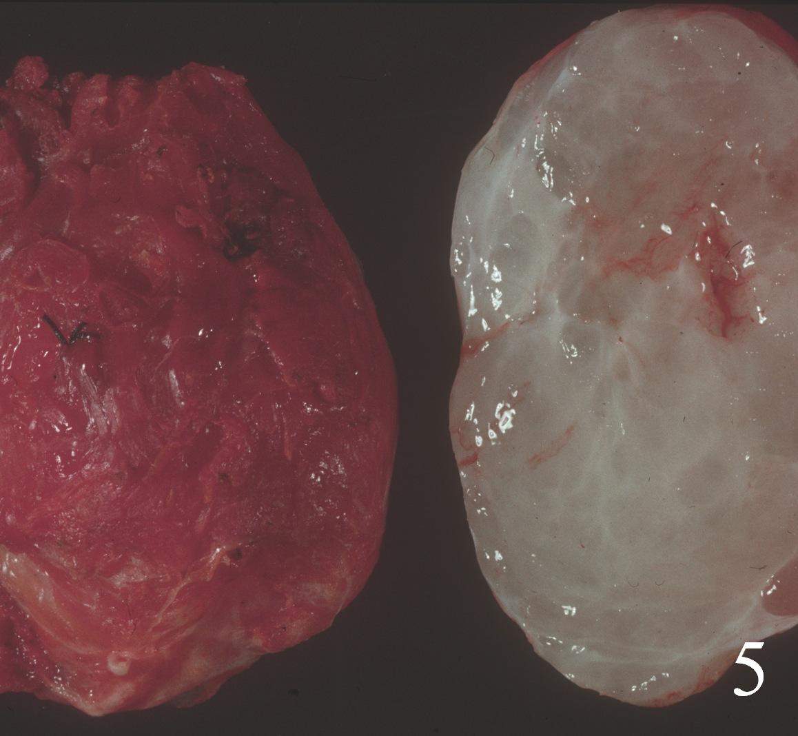 Fig. 5 Gross histology of myxoma showing a uniform gelatinous surface from the production of myxoid stroma
IMMUNOCHEMISTRY
Positive for spindle cells
Vimentin
CD 34 (50%)
Mucoid matrix
Alcian blue
Mucicarmine
Colloidal iron
Negative
S-100
Desmin
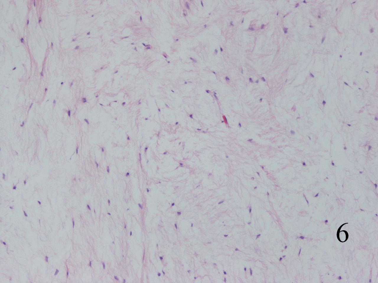
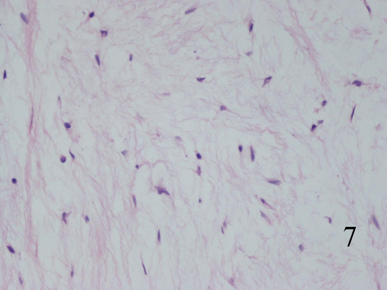 Fig. 6-7 Microscopic: Low power (Fig. 6) and high power magnification (Fig. 7) of a myxoma shows bland hypocellular uniform spindle shaped cells arranged in abundant myxoid stroma. There are no mitotic figures and no necrosis. The cells are very uniform.
PROGNOSIS
Biological behavior
Intramuscular myxoma
Rare local recurrence
No metastases
Recurring lesions are not destructive
Juxta-articular myxoma
Up to 30% rate of local recurrence---->infiltrative nature leads to recurrences
Even with complete excision
No metastasis
Digital myxoma
30% chance of local recurrence
No metastasis
TREATMENT
Excision, rarely recurs if complete excised
Cellular type appear to have same indolent behavior even after complete excision
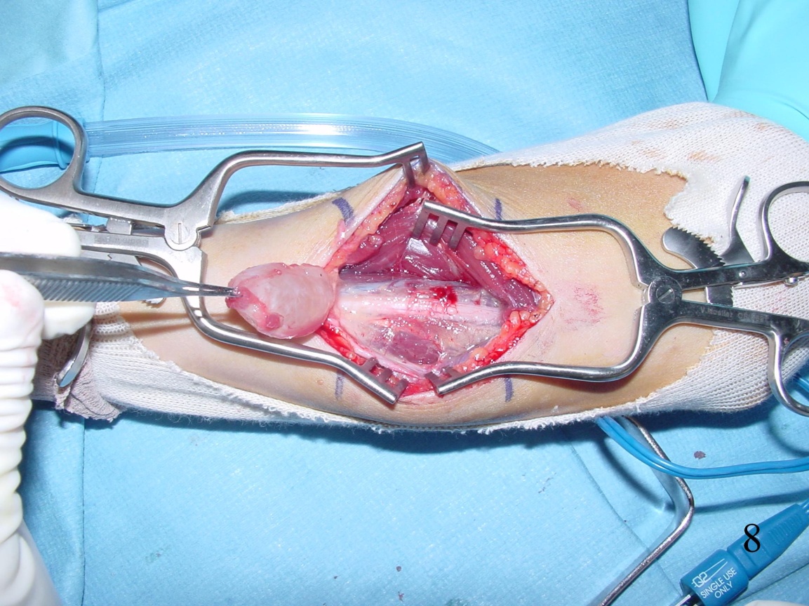 Fig. 8 Intraoperative photograph of resection of a myxoma from a forearm.
